Where is the kidney located?
The kidney is the retroperitoneal organ. To know where is the kidney located we must know the anatomy of kidney. The closely packed structure and numerous functions of the kidney illustrate the beautiful workmanship of our creator. It is not only applied to the kidney but to each and every part of our body.
Kidneys are a pair of excretory organs situated on the posterior abdominal wall, one each side of the vertebral column, behind the peritoneum. They remove waste products of metabolism and excess of water and salts from the blood and maintain its pH. “where is the kidney located” questions answer is discussed in the main paragraph of this article.
Where is the kidney located
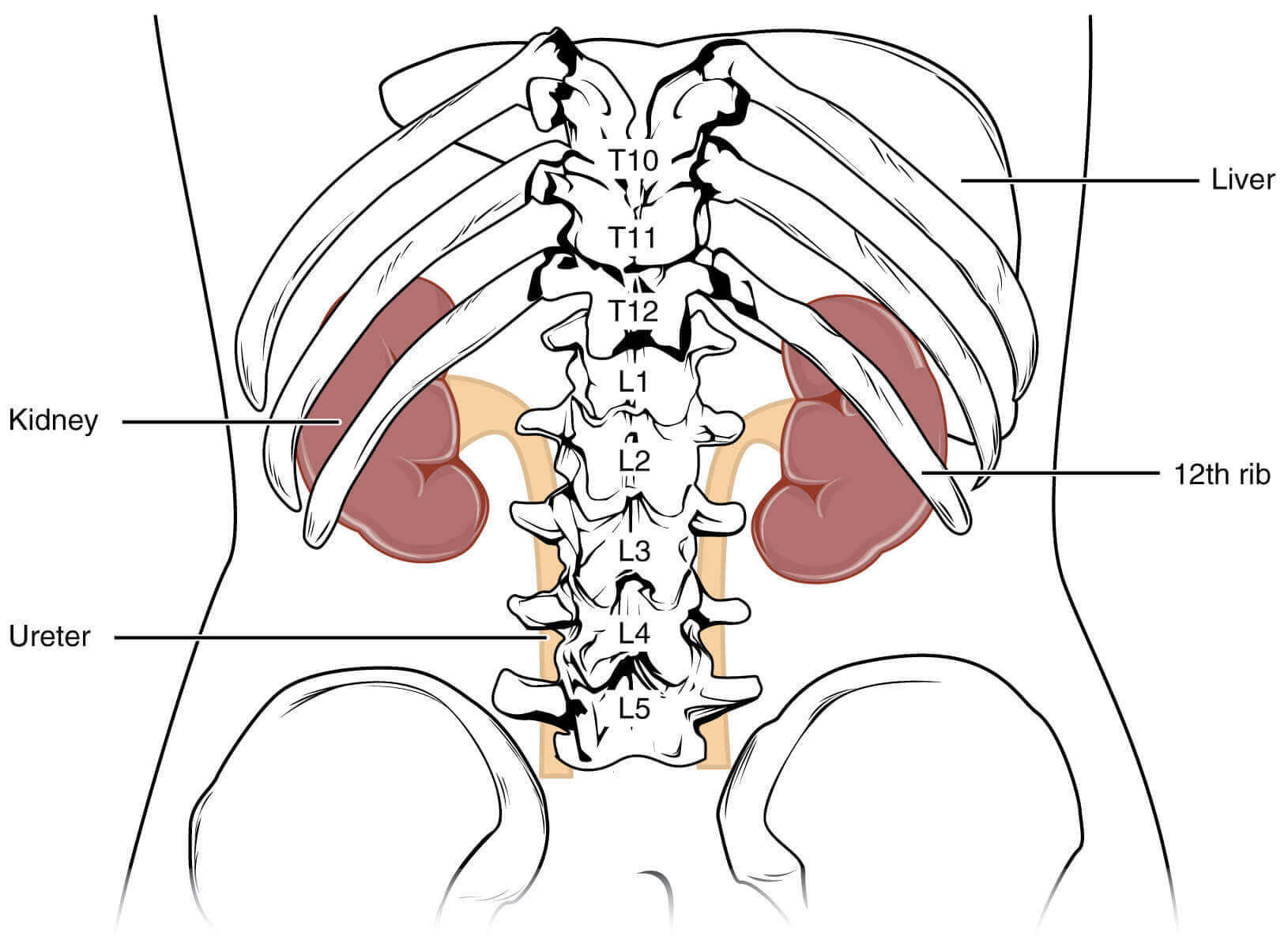
Let’s know about where is the kidney located.The kidneys occupy the epigastric, hypochondriac, lumbar and umbilical regions. Vertically they extend from the upper border of twelfth thoracic vertebrae. The right kidney is slightly lower than the left kidney, and the left kidney is a little nearer to the median plane than the right.
The transpyloric plane passes through the upper part of the hilus of the right kidney, and through the lower part of the hilus of the left kidney.I think you now know about the where is the kidney located
Anatomy of kidney:
Anatomy of kidney deals with the structure, shape size and location of the kidney. It is very important to know about the anatomy of kidney to know where is the kidney located. Anatomy of kidney is necessary to know about kidney hostology. To know about an organ we must know about its anatomy . Lets learn about the anatomy of kidney
Shape, size, weight and orientation
The individual kidney is about 11cm long, 6cm broad, and 3 cm thick. The left kidney is a brief longer and scrimpy than the right kidney. On average, the kidney weighs 150gm in males and 135gm in females. The kidneys are reddish-brown in color.
The long axis of the kidney is directed downwards and laterally so that upper poles are nearer to the median plane than the lower poles. The transverse axis is directed laterally and backward.
In fetus, the kidney is lobulated and is made up of about 12 lobules. After birth lobules gradually fuse, so that in adults the kidney is uniformly smooth. However, evidence of fetal lobulation may persist.
External features
Each kidney is bean-shaped. The kidney has upper and lower poles, medial and lateral borders, and anterior and posterior surfaces.
Two poles of the kidney
The upper pole is broad and is close contact with the corresponding suprarenal gland. The lower pole is pointed.
Two surfaces
The anterior surface is said to be irregular and the posterior surface flat, but it is often difficult to recognize the anterior and posterior aspects of the kidney by looking at the surfaces. The proper way to do this is to examine the structures present in the hilum as described below.
Two Borders
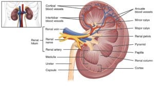
The lateral border is convex. The medial border is concave. Its middle part shows a depression, the hilus or hilum,.
Hilum
The following structures are seen in the hilum from the anterior to the posterior side.
- The renal vein
- The renal artery, and
- The renal pelvis, which is the expanded upper end of the ureter.
Examinations of these structures enable the anterior and posterior aspects of the kidney to be distinguished from each other. As the pelvis is continuous inferiorly with the ureter, the superior and inferior poles of the kidney can also be distinguished by examining the hilum. So it is possible to determine the side to which a kidney belongs by examining the structures in the hilum. Commonly, one of the branches of the renal artery enters the hilus behind the renal pelvis, and a tributary of the renal vein may be found in the same plane.
Relations of the kidneys
The kidneys are retroperitoneal organs and are only partly covered by the peritoneum anteriorly.
Relations common to the two kidneys
- The upper pole of each kidney is related to the corresponding suprarenal gland. The lower poles lie about 2.5 cm above the iliac crests.
- The medial border of each kidney is affiliated to:
- The suprarenal gland, above the hilus, and
- To the ureter below the hilus.
3. Posterior relations: the posterior surfaces of both kidneys are related to the following:
- diaphragm
- Medial and lateral arcuate ligaments
- Psoas major
- Quadratus lumorum
- Transerversus abdomonis
- Subcostal vessels; and
- Subcostal, iliohypogastric and ilioinguinal nerves
In addition, the right kidney is related to twelfth rib, and the left kidney to eleventh and twelveth ribs.
- The structures related to the hilum have been described earlier.
Other relations of the right kidney
Anterior relations
- Right suprarenal gland
- Liver
- The second part of the duodenum
- Hepatic flexure of the colon
- Small intestine
Out of these the hepatic and intestinal surfaces are covered by peritoneum.
The lateral border of the right kidney is related to the right lobe of the liver and to the hepatic flexure of the colon.
Other Relations of the left kidney
Anterior relations
- Left suprarenal glands
- Spleen
- Stomach
- Pancreas
- Splenic flexures and descending colon
- Jejunum
Out of these the gastric, splenic and jejunal surfaces are covered by peritoneum.
The lateral border of the left kidney is related to the spleen and to the descending colon.
Nerve supply
The kidney is supplied by the renal plexus, an offshoot of the coeliac plexus. It contains sympathetic (T10-L1) fibres which are chiefly vasomotor. The afferent nerves of the kidney belong to segments T10 to T12.
Exposure of the kidney from behind
In exposing the kidney from behind, the following layers have to be reflected one by one.
- Skin
- Superficial fascia
- The posterior layer of thoracolumbar fascia with lastissimus dorsi and serratus posterior inferior
- Erector spinae, which can be removed for convenience
- The middle layer of thoracolumbar fascia
- Quadratus lumborum
- The anterior layer of thoracolumbar fascia in which the related nerves are embedded.
Kidney Histology
Let’s know about the kidney histology. Kidney histology is importat to know. Kidney is covered by the capsule.The cortex of kidney shows cut sections of glomeruli, many sections of the proximal convoluted tubule, some sections of the distal convoluted tubule and few collecting ducts.
Sections through the pyramid of the medulla show light staining collecting ducts, sections of the loop of Henle, thick and thin segments of descending and ascending limbs, capillaries and connective tissue. So, this is the kidney histology. After reading this i think you have known some what abou thet kidney histology.
Nephron anatomy:
Nephron is a structural and functional unit of kidney. It is U-shaped tube which has a length of 50-55 mm long. It consist of a dilated blind end called Bowman’s Capsule and other open end called the renal tubule.
Takeway:
Kidneys are a pair of excretory organs situated on the posterior abdominal wall, one each side of the vertebral column, behind the peritoneum.The kidneys occupy the epigastric, hypochondriac, lumbar and umbilical regions.
I think now you know about the where is the kidney located.Kidney histology is importat to know. Kidey is covered by the capsule.The cortex of kidney shows cut sections of glomeruli, many sections of the proximal convoluted tubule, some sections of the distal convoluted tubule and few collecting ducts.
Do read: How to prevent kidney stones?