Anatomy of the stomach
Definition I Location I shape & size I capacity I External features I Blood supply I lymphatics drainage I Nerve supply I interior of the stomach I functions of the stomach I Histology
The gross anatomy of the stomach
The stomach is also called the gaster (Greek belly) or venter from which we have the adjective gastric applied to structure related to the organ. Let’s learn more about the anatomy of the stomach.
Definition of the stomach
The stomach is a muscular duffel forming the widest and most distensible part of the digestive tube. It is connected above to the lower end of the oesophagus and below to the duodenum. It acts as a reservoir of food and helps in digestion of carbohydrates, proteins and fats.
Location
The stomach is placed obliquely in the upper and left part of the abdomen occupying the epigastric, umbilical and left hypochondriac regions. Most of it lies beneath the cover of the left costal margin and the ribs
Shape
The shape of the stomach grounds upon the degree of its distension and that of the surrounding viscera e.g the colon. When abandoned, the stomach is somewhat J-shaped (vertical); when partially distended, It becomes pyriform in shape. In obese persons, it is more horizontal.
The shape of the stomach can be deliberated in the living by radiographic examination after giving a barium meal.
Size and Capacity
The stomach is a very distensible organ. It is about 25cm long and the mean capacity is one ounce (30ml) at birth, one litre (1000ml) at puberty and 11/2 to 2 litres or more in adults.
External features
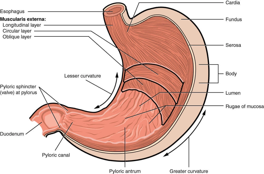
The anatomy of the stomach mainly begins with the external features of the stomach. The stomach annexe two orifices or openings, two curvatures or borders and two surfaces.
Two Offices
The cardiac orifice is joined by the lower end of the oesophagus. It lies behind the left 7th costal cartilage 2.5 cm from its junction with the sternum at the level of vertebra T11. There is physiological evidence of sphincteric action at the side but a sphincter cannot be demonstrated anatomically.
The pyloric orifice opens into the duodenum. In an empty stomach and in the supine position, it lies 1.2 cm to the right of a median plane at the level of the lower border of vertebra L1 or transpyloric plane. Its position is indicated on the surface of the stomach.
- By a circular groove (pyloric constriction, i,e. sthenic. Sometimes it may be hypersthenic (thin and long) hypersthenic pylorus) produced by the underlying pyloric sphincter or pylorus (pylorus = gate guard) which feels like a large firm nodule.
- By the prepyloric vein of Mayo which lies in front of the constriction.
Two Curvatures
The lesser curvature is concave and forms the right border of the stomach. It provides attachment to the lesser omentum. The most dependent part of the curvature is marked by the angular notch or incisura angularis.
The greater curvature is convex and constitutes the left border of the stomach. It provides an adapter to the greater omentum, the gastrosplenic ligament and the gastrophrenic ligament. At its upper end, the greater curvature presents the cardiac notch which separates it from the oesophagus. It is 5 times longer than lesser curvature.
Two Surfaces
- The anterior or anterosuperior surface faces forwards and upwards.
- The posterior or posteroinferior surface faces backwards and downwards.
Two parts Subdivided into Four
The stomach is divided into two parts.
- Cardiac and
- Pyloric,
by a line, is drawn downwards and to the left from the incisura angularis. The larger cardiac part is further subdivided into the fundus and body and the smaller pyloric part is subdivided into the pyloric antrum and pyloric canal.

Cardiac Part
let’s begin anatomy of the stomach with cardiac part.
- The fundus of the stomach is the upper convex dome-shaped part situated above a horizontal line drawn at the level of the cardiac orifice. It is commonly distended with gas which is seen clearly in radiographic examination under the left dome of the diaphragm.
- The body of the stomach lies between the fundus and the pyloric antrum. It can be distended enormously along the greater curvature. The gastric glands distributed in the fundus and body of the stomach contain all three type of secretary cells namely
- The mucous cells.
- The chief peptic or zymogenic cells which secrete the digestive enzymes.
- The parietal or oxyntic cells which secrete HCL.
Pyloric Part
- The pyloric antrum is separated from the pyloric canal by an inconstant sulcus, sulcus intermedius present on the greater curvature. It is about 7.5 cm long. The pyloric glands are richest in mucous cells.
- The pyloric canal is about 2.5 cm long. It is narrow and tubular. At its right end, it terminates at the pylorus.
Peritoneal Relations
The stomach is lined by peritoneum on both its surfaces. At the lesser curvature, the layers of peritoneum lining the anterior and posterior surface meet and become continuous with the lesser omentum.
Along the greater part of the greater curvature, the two layers meet to from the greater omentum.
Near the fundus, the two layers meet to form the gastrosplenic ligament.
Near the cardiac end, the peritoneum on the posterior surface is reflected on to the diaphragm as the gastrophrenic ligament. Cranial to this ligament a small part of the posterior surface of the stomach is in direct contact with the diaphragm (left crus). This is the bare area of the stomach. The greater and lesser curvatures along the peritoneal reflections are also bare.
Visceral Reactions
The anterior surface of the stomach is related to the liver, the diaphragm, transverse colon and the anterior abdominal wall. The area of stomach related to these structures. The diaphragm separates the stomach from the left pleura, the pericardium and the sixth to ninth ribs. The costal cartilages are separated from the stomach by the transversus abdominis. Gastric nerves and vessels ramify deep to the peritoneum.
The space between the left costal margin and lower edge of left lung on the stomach is known as Traube’s space. Normally, on percussion there is resonant note over this space; but in splenomegaly or pleural effusion, a dull note is felt at this site.
The posterior surface of the stomach is related to structures forming the stomach bed, all of which are separated from the stomach by the cavity of the lesser sac.
The structures are:
- Diaphragm
- Left kidney
- Left suprarenal gland
- Pancreas
- Transverse mesocolon
- Splenic flexure of the colon
- Splenic artery (19.9). Sometimes the spleen is also included in the stomach bed, but it is also included in the stomach by the cavity of the greater sac (and not of the lesser sac). Gastric nerves and vessels ramify deep to the peritoneum.
Blood Supply
The stomach is supplied along the lesser curvature by:
- The left gastric artery, a branch of the celiac trunk.
- The right gastric artery, a branch of the proper hepatic artery.
- Along the greater curvature, it is supplied by the right gastroepiploic artery, a branch of the gastroduodenal.
- The left gastroepiploic artery, an arm of the splenic.
- The fundus is supplied by 5 to 7 short gastric arteries, which are also branches of the splenic artery

The veins of the stomach drain into the portal superior mesenteric and splenic veins. Right and left a gastric drain in the portal vein. Right gastroepiploic and short gastric veins terminate in the splenic vein
Lymphatic drainage
The anatomy of the stomach can be asunder into four lymphatic territories. The drainage of these areas is as followers.
Area, i.e. the upper part of left 1/3rd drains into the pancreaticosplenic nodes lying along the splenic artery, i.g. on the back of the stomach. Lymph vessels from these nodes travel along the splenic artery to reach the celiac nodes.
Area, i.e. right 2/3rd drains into the left gastric nodes lying along the artery of the same name. These nodes also conduit the abdominal part of the oesophagus. Lymph from these nodes drains into the coeliac nodes.
Area, i.e. the lower part of left 1/3rd drains into the right gastroepiploic nodes that lie along the artery of the same name. Lymph vessels arising in these nodes drain into the sub pyloric nodes which lie in the angle between the first and second part of the duodenum. From here the lymph is drained further into the hepatic nodes that lie along the hepatic artery; and finally into the coeliac nodes.
Lymph from an area, i.g. pyloric part drains in different directions into the pyloric, hepatic and left gastric node to the coeliac nodes. Note the manner in which the organ is subdivided into a to d different territories gastric nodes and passes from all these nodes to the coeliac nodes. Note that lymph from all areas of the stomach ultimately reaches the coeliac nodes. From here it passes through the intestinal lymph trunk to reach the cistern cliyli
Nerve Supply
The stomach is supplied by sympathetic and parasympathetic nerves. The sympathetic nerves are derived from thoracic six to ten segments of the spinal cord, via the greater splanchnic nerves and coeliac and hepatic plexuses. These nerves are:
- Vasomotor.
- Motor to the pyloric sphincter, but inhibitory to the rest of pyloric sphincter, but inhibitory to the rest of the gastric musculature.
- The chief pathway for pain sensation from the stomach.
The parasympathetic nerves (19.12 a & b) are derived from the vagi, through the oesophageal plexus and gastric nerves. The anterior gastric never (made up one or two trunks) contains mainly the left vagal fibres and the posterior gastric nerve (again made up of one to two trunks) contains mainly the right vagal fibres.
Nerve supply of the stomach:
(a) Anterior gastric nerve and
(b) The posterior gastric nerve.
The anterior gastric nerve divides into:
- A number of gastric branches for the anterior surface of the fundus and body of the stomach.
- Two pyloric branches, one for the pyloric antrum and another for the pylorus.
The posterior gastric nerve divides into:
- Smaller, gastric branches for the posterior surface of the fundus, the body and the pyloric antrum.
- Larger, coeliac branches for the coeliac plexus. Parasympathetic nerves are motor and secretomotor to the stomach. Their stimulation causes increased motility of the stomach and secretion of gastric juice rich in pepsin and HCL. These are inhibitory to the pyloric sphincter.
Interior of the stomach
The anatomy of the stomach also deals with the interior of the stomach. Open the stomach along the greater curvature and examine the mucous membrane with a hand lens. Then strip the mucous membrane from one part and expose the internal muscle coat. Dissect the muscle coat, e.g. outer longitudinal middle circular and inner oblique muscle fibres. Feel thickened pyloric sphincter. Incise the beginning of duodenum and examine the duodenal and pyloric aspects of the pyloric sphincter.
features
The stomach has to be incised to see its internal structure.
- The mucosa of an empty stomach is thrown into folds termed as gastric rugae. The rugae are longitudinal along the lesser curvature and are irregular elsewhere. The rugae are flattened in a distended stomach. On the mucosal covering, there are numerous miniature depressions that can be seen with a hand lens. These are the gastric pits. The gastric glands open into these pits.
The part of the lumen of the stomach that lies along the lesser curvature and has longitudinal rugae is called the gastric canal or magenstrasse. This canal allows rapid passage of swallowed liquids along the lesser curvature directly to the lower part before it spreads to the other part of the stomach. Thus lesser curvature bears maximum insult of the swallowed liquid, which makes it vulnerable to peptic ulcer. So, beware of your drinks.
- The submucous coat is made of connective tissue, arterioles and nerve plexus.
- Muscle coat is arranged as under:
- Longitudinal fibres are most superficial mainly along the curvatures.
- Inner circular fibres encircle the body and
- The deepest layer consists of oblique fibres which loop over the cardiac notch. Some fibres spread in the fundus and body of the stomach. Rest form a well-developed ridge on each side of the lesser curvature. These fibres on contraction form “gastric canal” for the passage of fluids.
- The serous coat consists of the peritoneal covering.
Functions of Stomach
- The stomach acts primarily as a reservoir of food. It also acts as a mixer of food.
- By its peristaltic movements, it softens and mixes the food with the gastric juices.
- The gastric glands produce the gastric juice which contains enzymes that play an important role in the digestion of food.
- The gastric glands also produce hydrochloric acid which destroys many organisms present in food and drink.
- The lining cells of the stomach produce abundant mucus which protects the gastric mucosa against the corrosive action of hydrochloric acid.
- Some substances like alcohol, water, salt and few drugs are absorbed in the stomach.
- The stomach produces the “intrinsic factor” of Castle which helps in the absorption of vitamin B12.
Microscopic anatomy of the stomach
At the cardiac confine of the stomach, the stratified epithelium of oesophagus abruptly changes to simple columnar epithelium of the stomach.
Cardiac End
Mucous membrane: The epithelium is simple columnar with small tubular glands. Lower bisected of the gland is secretory and the upper half is the conducting part. Muscularis mucosae consist of smooth muscle fibres.
Sub Mucosa: It consists of loose connective tissue with Meissner’s (German histologist 1829-1909) plexus.
Muscularis Externa: It is made of outer longitudinal and an inner circular layer including the myenteric plexus of nerves or Auerbach’s plexus (German anatomist 1828-97).
Serosa: It is lined by a single layer of squamous cells.
Fundus and Body of Stomach
Mucous membrane: It contains tall simple tubular gastric glands. Upper one-third is conducting, while lower two-thirds is secretory. The various cell types conceive in the gland are chief or zymogenic, oxyntic or parietal and mucous neck cells
Muscularis mucosae and submucosa are same.
Muscularis Externa: It bottles up an additional innermost oblique coat of muscle fibres.
The serosa is same as of the cardiac end.
Pyloric part
Mucous membrane: There are pyloric glands which consist of basal one-third as mucous secretory component and upper two-third as conducting part. Muscularis mucosae are built of two layers of fibres. The submucosa is same as in the cardiac end.
Muscularis external comprises a thick layer of circular fibres forming the pyloric sphincter. The serosa is same as of the cardiac end and the anatomy of the stomach ends here.
Conclusion
In this gross anatomy of the stomach post, you have learned all the detail about the anatomy of the stomach with the microscopic finding.
It is the first intra-abdominal part of the gastrointestinal (GI) tract. It may take varying shapes, depending on the build and posture of the person and the state of fullness of the organ.
The stomach muscles contract systematically, churning food to embellish digestion.